Your spine is a marvel of nature’s engineering, a flexible column made of small bones, or vertebrae, which houses the spinal cord – a crucial highway for messages between your brain and the rest of your body. When something goes wrong in your cervical spine – the neck portion of your spine – it can cause severe neck pain and disrupt your daily life.
Cervical laminoplasty, a surgical procedure performed on the neck, can bring significant relief to individuals experiencing pain and discomfort due to spinal cord or nerve pressure. In a study of thirty-two patients, researchers found that all cervical laminoplasty using autograft patients reported less pain three months and after two years 91% reported improvements as well.
“Cervical laminoplasty is a non-fusion, decompression procedure for cervical spondylotic myelopathy (CSM). It is most commonly indicated for patients with multilevel stenosis who have preserved sagittal alignment and minimal to no axial neck pain related to spondylosis.”
Weinberg DS, Rhee JM. Cervical laminoplasty: indication, technique, complications. J Spine Surg. 2020 Mar;6(1):290-301. doi: 10.21037/jss.2020.01.05. PMID: 32309667; PMCID: PMC7154346.
When is Cervical Laminoplasty Recommended?
Cervical laminoplasty is typically recommended in a few specific situations. If you’re dealing with spinal cord compression, also known as cervical myelopathy, due to conditions like cervical stenosis or degenerative disc disease, and it’s causing you symptoms such as chronic neck pain, weakness, numbness, or tingling in your arms or hands, and conservative treatments such as physical therapy are ineffective, a cervical laminoplasty may be recommended.
Cervical Laminoplasty Surgical Procedure
Before the procedure, an intravenous line, or IV, will be initiated, possibly delivering antibiotics to prevent infection. General anesthesia ensures that you will remain unconscious and pain-free throughout the operation.
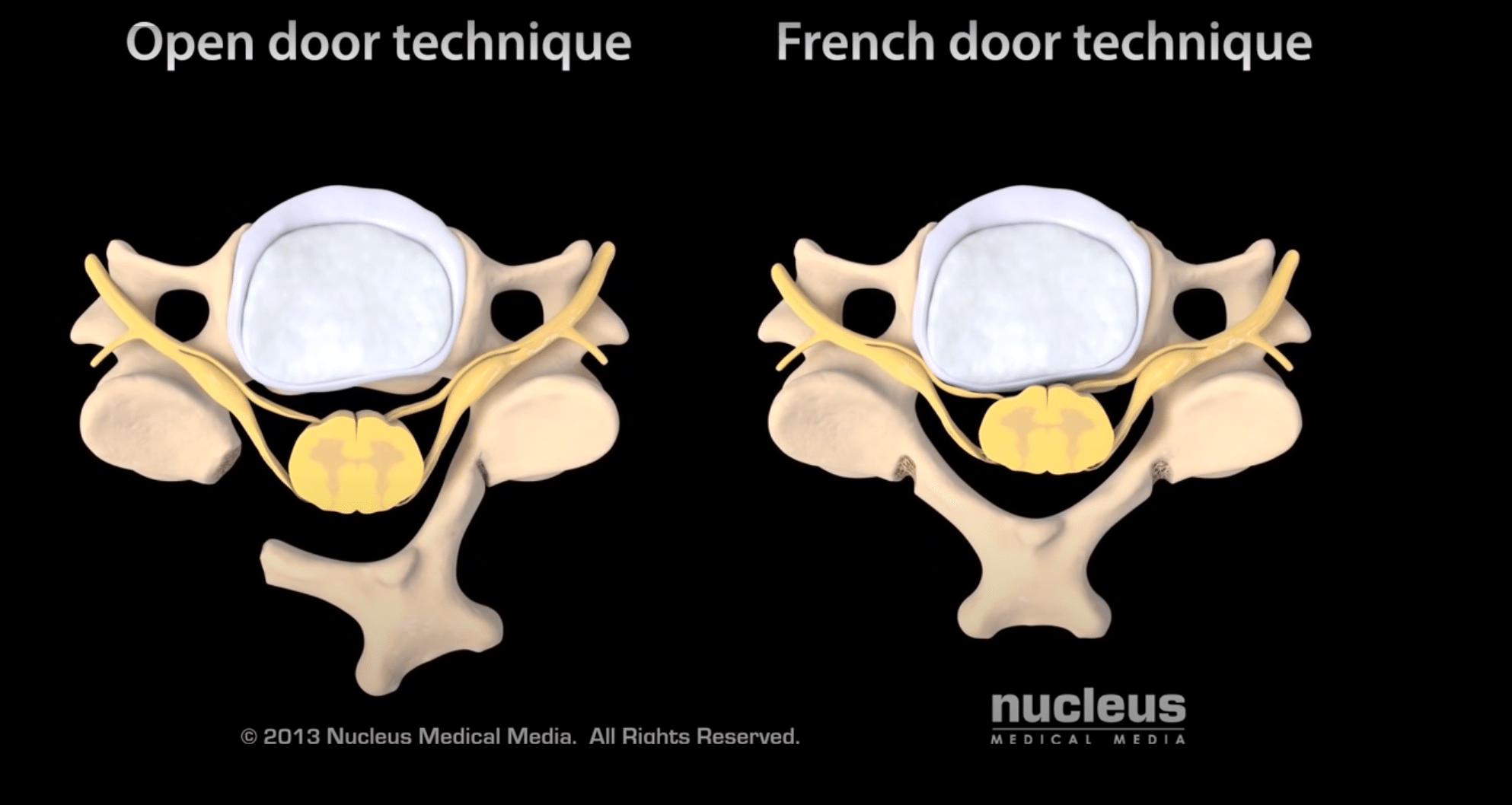
During the surgery, your surgeon will make an incision on the back of your neck, revealing the laminae of the affected vertebrae. The surgeon will remove the outer layer of bone from each lamina, forming two troughs. The vertebral arch is then pulled open, relieving pressure on the spinal cord.
There are two techniques commonly used during this procedure: the open door and the French door, or double door, technique. The former involves cutting through one trough and using the other as a hinge, while the latter uses both troughs as hinges, allowing the surgeon to split the spinous process and open the vertebral arch in the middle.
Usually, the vertebral arch will be opened on more than one of the cervical vertebrae. Bone graft material, secured by metal plates, may be inserted into each vertebral arch to maintain its open position.
Once the procedure is complete, the surgeon will close your incision with sutures, surgical skin glue, or staples. Your neck will be placed in a collar to promote healing and prevent unnecessary movement.
Recovery Post-Surgery
Following your procedure, you will be moved to the recovery area for monitoring. Pain medication will be provided as necessary. You will wear a neck collar for several weeks to facilitate proper healing.
Typically, patients are discharged from the hospital within two to three days post-surgery. The road to recovery varies for each patient, and it’s important to follow your doctor’s guidelines for the best outcome.
