The human spine is a remarkable structure that serves as both a support system and a conduit for the nervous system. Within its complex anatomy, several canals play crucial roles in protecting and facilitating the function of the spinal cord and its associated nerves. Among these, the central canal and neural foramen stand out as key features that ensure the proper functioning of the body’s communication network.
The Central Canal: A Pathway for the Spinal Cord
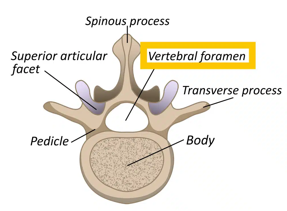
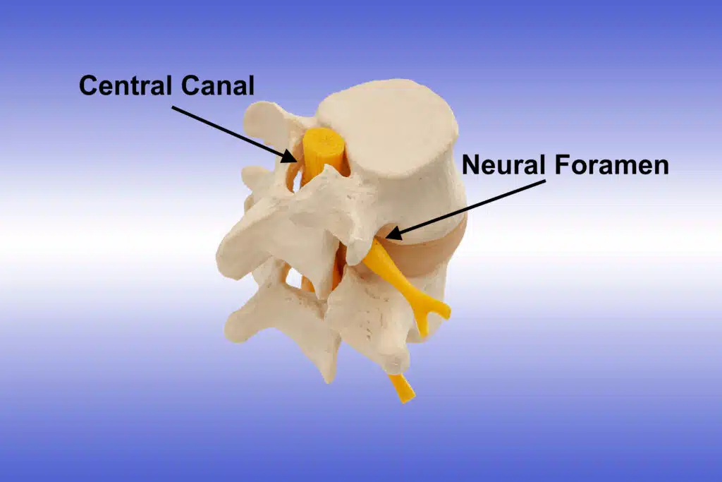
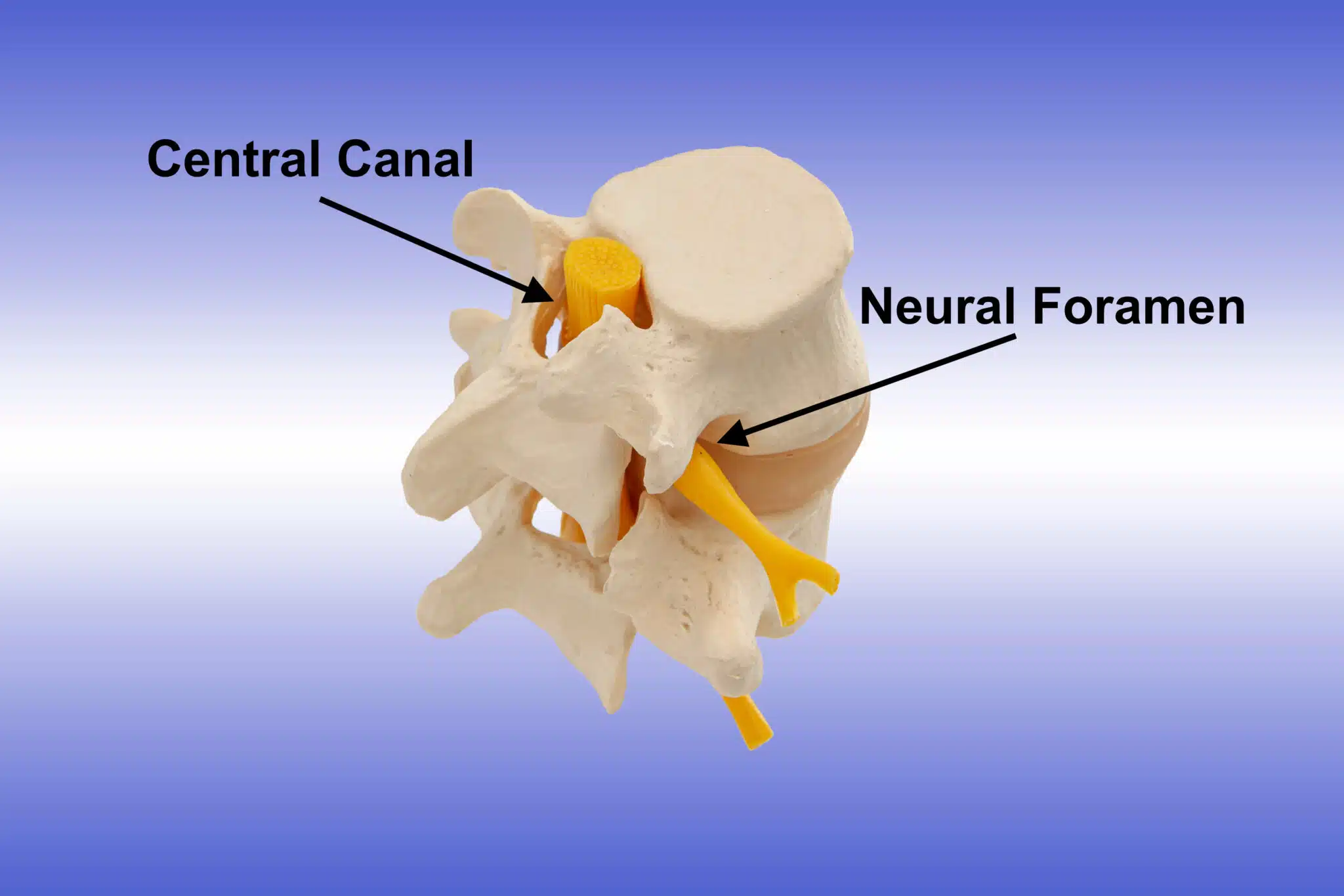
The central canal, also known as the central spinal canal or the vertebral canal, is a cylindrical channel that runs the length of the spinal cord. It is located at the center of the vertebral column and is surrounded by the vertebral arches, which are bony structures that form the spinal column’s protective framework. The primary role of the central canal is to house and protect the spinal cord, a vital component of the central nervous system.

Structure and Function
The central canal is lined with a delicate layer of cells known as ependymal cells. These cells are responsible for producing cerebrospinal fluid (CSF), a clear and nutrient-rich fluid that cushions and supports the spinal cord. CSF helps maintain a stable environment for the spinal cord by providing protection against mechanical shocks and maintaining a consistent pressure.
While the central canal serves as a conduit for CSF circulation, its importance goes beyond mere fluid dynamics. The canal’s presence also enables communication between different levels of the spinal cord, facilitating the exchange of signals that control movement, sensation, and various bodily functions.
Neural Foramen: Gateways for Spinal Nerves
Neural foramina (plural of foramen) are small openings located on either side of the vertebral column. These foramina serve as exit points for spinal nerves, allowing them to extend outwards from the spinal cord and connect with other parts of the body. Each spinal nerve, except for the first cervical nerve, exits the vertebral column through a pair of neural foramina—one on the left side and another on the right.

Structure and Function
Neural foramina are formed by the alignment of adjacent vertebrae and their associated structures, including the vertebral body, pedicles, and laminae. These bony elements create a space through which spinal nerves can pass without being compressed or impeded. The neural foramina are integral to the transmission of nerve signals between the central nervous system and the peripheral nervous system.
The health and functionality of the neural foramina are crucial to ensure that spinal nerves can exit the spinal cord without obstruction. Any condition that narrows or compresses the neural foramina can lead to nerve impingement, resulting in symptoms such as pain, numbness, tingling, or weakness in the areas served by the affected nerves. Common conditions that can affect the neural foramina include herniated discs, bone spurs, and spinal stenosis.
Conclusion
The canals of the spine—the central canal and neural foramina—play essential roles in maintaining the health and functionality of the nervous system. The central canal protects the spinal cord and facilitates the circulation of cerebrospinal fluid, while neural foramina provide exit points for spinal nerves to connect with the rest of the body. Any condition that narrows or compresses the these canals can lead to nerve impingement, resulting in symptoms such as pain, numbness, tingling, or weakness in the areas served by the affected nerves.
