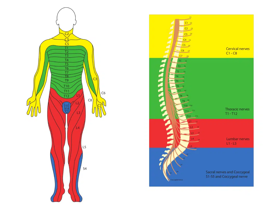
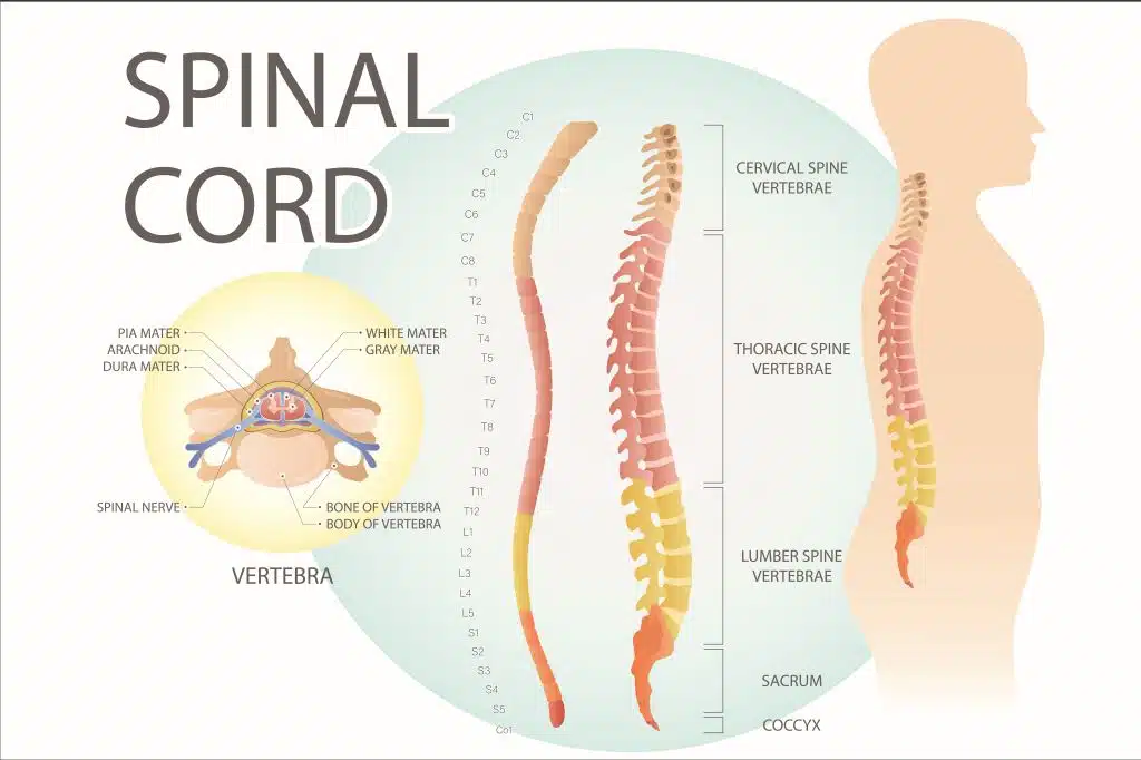
The spinal cord is a long, thin, cylindrical structure that runs from the base of the brain down through the vertebral column (spine or backbone) and terminates near the bottom of the lower back. It is part of the central nervous system (CNS) and is responsible for transmitting signals between the brain and the rest of the body.
The spinal cord is made up of millions of nerve fibers, which are bundled into tracts that carry different types of information. Some tracts carry signals related to sensory information such as touch, temperature, and pain, while others carry signals related to movement and coordination of muscles.
How does the spinal cord work?
The spinal cord works by transmitting signals between the brain and the rest of the body. It receives sensory information from the body and sends it to the brain for processing, and it also sends motor signals from the brain to the muscles to control movement.
When a sensory signal is detected in the body, it travels along sensory neurons to the spinal cord, where it is processed and relayed to the brain. The brain then sends back a motor signal along motor neurons, which travel from the brain down through the spinal cord and out to the muscles to control movement.
The spinal cord also contains reflex pathways, which allow for rapid, automatic responses to certain stimuli without the need for input from the brain. For example, when you touch something hot, the sensory information travels to the spinal cord and triggers a reflex that causes you to quickly withdraw your hand without consciously thinking about it.
What are the regions of the spinal cord? What is their function?
Different parts of the spinal cord are responsible for controlling different functions in the body. Generally, the higher up the spinal cord the injury or damage occurs, the greater the impact it will have on bodily functions. Here’s a brief overview of the functions controlled by different parts of the spinal cord:
- Cervical region: The cervical region of the spinal cord controls the movements and sensations of the neck, shoulders, arms, and hands. Injuries to this region can cause paralysis or weakness in these areas, as well as difficulty breathing and speaking.
- Thoracic region: The thoracic region of the spinal cord controls the movement and sensation of the chest, mid-back, and abdominal muscles. Injuries to this region can cause difficulty with breathing and coughing, and may also affect trunk stability and bowel and bladder control.
- Lumbar region: The lumbar region of the spinal cord controls the movement and sensation of the hips, legs, and feet. Injuries to this region can cause paralysis or weakness in the legs, as well as difficulty with bowel and bladder control.
At the lower end of the lumbar region of the spinal cord, the cord loses its cylindrical structure and the nerves split off. This region is known as the “cauda equina”, and carries the lower lumbar and sacral nerve roots.
What is the cauda equina?
The cauda equina is a bundle of nerve roots that originates from the lower end of the spinal cord and extends down through the lower back and into the pelvis. The term “cauda equina” means “horse’s tail” in Latin, and the bundle of nerves is so named because of its resemblance to a horse’s tail.
The cauda equina is made up of many individual nerve roots, which emerge from the spinal cord and pass through small openings in the vertebrae of the lower back. These nerve roots then continue downward and form a network of nerves that control the sensation and movement of the legs, feet, and pelvic organs.
Damage to the cauda equina can cause a variety of symptoms, including lower back pain, leg pain, numbness or tingling in the legs or feet, weakness in the legs, and problems with bladder or bowel control. This condition is known as cauda equina syndrome and is a medical emergency that requires immediate treatment to prevent permanent damage to the nerves.
Nerve roots
Spinal nerve roots are bundles of nerve fibers that emerge from the spinal cord and exit the vertebral column through small openings between adjacent vertebrae. There are 31 pairs of nerve roots in the human body, each of which is composed of a sensory nerve root and a motor nerve root.
The sensory nerve root contains nerve fibers that transmit information from the body to the spinal cord, such as sensations of touch, pain, and temperature. The motor nerve root contains nerve fibers that transmit information from the spinal cord to various muscles, controlling voluntary movement.
The nerve roots are numbered based on their location in the spinal cord, with the first cervical nerve root (C1) located at the top of the spinal cord near the base of the brain, and the last sacral nerve root (S5) located at the bottom. Each pair of nerve roots is associated with a dermatome, which is an area of skin that is supplied with sensory fibers from a single spinal nerve root, and a myotome, which is a group of muscles supplied by a single spinal nerve root.

Damage or compression of a nerve root can cause pain, weakness, numbness, or tingling in the area of the body supplied by that nerve root. Nerve root compression is a common cause of conditions such as herniated discs, spinal stenosis, and sciatica. Treatment for nerve root compression may include medication, physical therapy, spinal steroid injections, and in severe cases, surgery to relieve pressure on the affected nerve root.
