Summary
A transitional vertebra is a common variation found in the human spine where a vertebral segment possesses characteristics that appear to bridge two adjacent regions of the spine. Transitional vertebrae are typically congenital, meaning they are present from birth, and usually do not cause any symptoms. Rarely, they can cause increased stress on adjacent vertebrae, nerve compression, and abnormal spinal curvatures.
Understanding Transitional Vertebrae
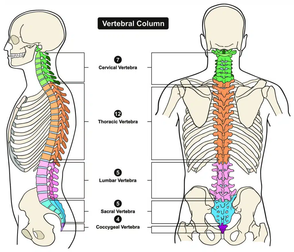
In the normal human spine, there are 33 vertebrae, grouped into five regions: cervical (7), thoracic (12), lumbar (5), sacral (5-fused), and coccygeal (4-fused). However, sometimes, a vertebra may demonstrate characteristics that appear to bridge two adjacent regions. This unique vertebra, known as a transitional vertebra, possesses a combination of features from the neighboring vertebral regions.
A transitional vertebra is also known as a transitional segment or transitional anomaly.

Causes of Transitional Vertebrae
Transitional vertebrae are typically congenital, meaning they are present from birth. Their occurrence results from a developmental anomaly during the early stages of fetal growth. The specific cause of this anomaly is not always clear, but it may be influenced by genetic factors and environmental influences during embryonic development.
Types of Transitional Vertebrae
The most common location for a transitional vertebra is in the lumbosacral junction, where the lumbar spine (lower back) meets the sacrum (triangular bone at the base of the spine). In this area, the vertebra may exhibit characteristics of both lumbar and sacral vertebrae.
There are several types of transitional vertebrae, each characterized by its unique features and potential effects on the spine:
- Lumbarization of the Sacrum: In this type, a lumbar vertebra retains its individuality and fails to fuse with the rest of the sacrum, appearing as an additional lumbar-like vertebra.
- Sacralization of the Lumbar: In this type, the first sacral vertebra fuses with the last lumbar vertebra, causing the lumbar region to appear as if it has one fewer vertebra.
- Cervical Rib: A less common type of transitional vertebra, where an extra rib-like bony projection extends from one of the cervical vertebrae (usually the seventh).
Prevelance
Transitional vertebrae are relatively common with studies indicating an occurrence rate ranging from 4% to 30% among the population (Konin 2010).
Implications and Clinical Significance
Most individuals with a transitional vertebra remain asymptomatic and may never even be aware of its presence. However, in some cases, the transitional vertebra can lead to certain spinal conditions and issues:
- Increased Stress on Adjacent Vertebrae: The presence of a transitional vertebra can alter the biomechanics of the spine, potentially leading to increased stress on neighboring vertebrae. This may contribute to the development of spinal conditions such as spondylolisthesis, where one vertebra slips over another.
- Nerve Compression: Rarely, a transitional vertebra may cause compression of nearby nerves, resulting in pain, tingling, or weakness in the legs.
- Abnormal Spinal Curvatures: In some cases, a transitional vertebra can contribute to the development of abnormal spinal curvatures, such as scoliosis or kyphosis.
Management and Treatment
Treatment for a transitional vertebra is typically only necessary if it causes significant symptoms or complications. Conservative approaches, such as physical therapy, pain management, and lifestyle modifications, are usually the first line of treatment. Surgical intervention is considered in severe cases where nerve compression or significant instability is present.
Takeaways
Transitional vertebrae are unique variations found in the human spine that possess characteristics bridging two adjacent regions. Typically congenital, these anomalies result from developmental anomalies during fetal growth, and their most common location is in the lumbosacral junction. There are several types of transitional vertebrae, including lumbarization of the sacrum, sacralization of the lumbar, and cervical rib. While most individuals with a transitional vertebra remain asymptomatic, in some cases, it can lead to spinal conditions and issues such as increased stress on adjacent vertebrae, nerve compression, and abnormal spinal curvatures. Treatment is usually conservative, with physical therapy and pain management, but surgery may be considered in severe cases. Understanding transitional vertebrae aids in providing appropriate care for affected individuals.
Sources
Konin GP, Walz DM. Lumbosacral transitional vertebrae: classification, imaging findings, and clinical relevance. AJNR Am J Neuroradiol. 2010;31(10):1778-1786